

Advanced bone regeneration techniques rebuild jawbone and periodontal structures lost to gum disease, tooth extraction, or trauma, using bone grafting materials, protective membranes, and precision imaging to create a stable foundation for your teeth or a future dental implant. The most important decision in regenerative care is not which material to use, but which technique fits the specific shape and location of your bone loss, because a vertical defect beside a natural tooth, an empty extraction socket, and a thin ridge under the sinus each call for a different approach.
At Advanced Periodontics of N.J., Dr. Peto and our clinical team use these techniques to help patients throughout River Edge and the surrounding Bergen County communities address bone loss that once meant limited treatment options. Our approach draws on evidence-based protocols across the full range of periodontal conditions we treat, from early bone loss to more advanced disease, and this guide walks through how we match each regenerative technique to the type of bone loss it treats best.
What Causes Bone Loss Around Teeth?
Bone loss around the teeth develops gradually and often without obvious symptoms until the supporting structure is already compromised. Chronic periodontal disease is the most common cause, since long-standing bacterial infection breaks down the ligament and bone that anchor teeth in the jaw. National survey data published by CDC researchers in the Journal of Dental Research (Eke and colleagues, 2012) found that 47.2% of U.S. adults aged 30 and older had some form of periodontitis, which is why bone loss is such a common finding at periodontal evaluations.
Tooth extraction is another frequent cause, as the bone that once surrounded a tooth root begins to resorb once that stimulation is gone. Facial trauma, ill-fitting dentures, and chronic infection around a failing dental implant can each contribute to further bone loss as well. Left unaddressed, this loss of bone can affect the stability of neighboring teeth, the fit of a denture, and the long-term success of a future dental implant. Recognizing these causes early gives our team more options for rebuilding lost bone before the damage becomes extensive, whether through membrane-guided techniques, bone grafting materials, or a combination of both.
Why the Shape of the Bone Defect Drives the Treatment Choice
Two patients can both be told they have “bone loss” and still need very different treatment. Periodontists describe bone loss by its pattern, because the pattern predicts how well new bone can be encouraged to grow back.
Vertical (angular) defects are narrow pockets of missing bone that run down the side of a single tooth root. When the defect still has bony walls on two or three sides, those walls act like a container that holds graft material and supplies the cells that form new bone, which makes these defects the most favorable for regeneration. Horizontal bone loss, by contrast, is an even drop in the bone level across several teeth. With no walls to contain a graft, rebuilding horizontal bone height around natural teeth is far less predictable, so treatment usually focuses on controlling infection and reducing pockets instead.
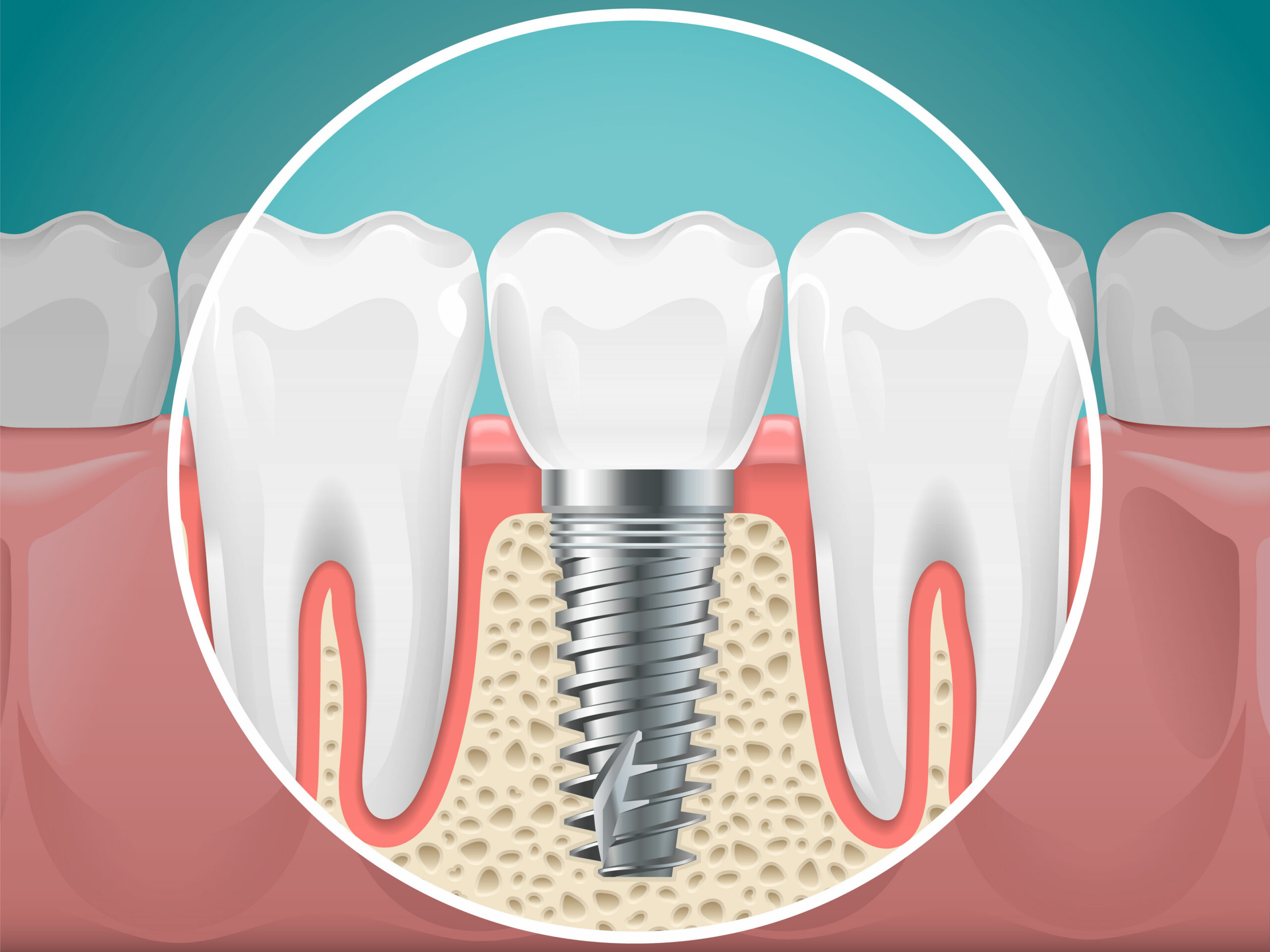
Bone loss also happens where there is no tooth at all. An extraction socket, a ridge that has narrowed after years without a tooth, and a thin layer of bone beneath the sinus in the upper back jaw are all “site development” problems, where the goal is building enough bone for an implant rather than saving a natural tooth.
Matching the Technique to the Type of Bone Loss
Once imaging and periodontal probing show the pattern of bone loss, the treatment options narrow considerably. The situations below are the ones we see most often.
A Vertical Defect Beside a Natural Tooth
For a contained defect next to a tooth worth saving, periodontal regenerative surgery is generally the first choice. The European Federation of Periodontology’s S3 level clinical practice guideline recommends regenerative surgery for teeth with residual deep pockets associated with intrabony defects 3 mm or deeper, using barrier membranes or enamel matrix derivative with or without graft material. Guided tissue regeneration is the best-known version of this approach, and our article on how guided tissue regeneration restores bone around teeth covers that procedure step by step.
Horizontal Bone Loss and Residual Pockets
When bone has dropped evenly across several teeth, the priority shifts to eliminating the deep pockets where bacteria collect. The same EFP guideline recommends repeating non-surgical cleaning for moderately deep residual pockets of 4 to 5 mm and considering access flap surgery for pockets of 6 mm or deeper. Depending on the case, that may mean osseous pocket reduction surgery or a laser-based protocol, with regenerative materials reserved for any individual sites that have a favorable defect shape.
An Empty Socket After an Extraction
The fastest bone loss most people ever experience happens in the months after a tooth is removed. Placing graft material into the socket at the time of extraction helps limit that collapse, and our article on why ridge preservation should happen immediately after extraction explains the timing in detail.
A Narrow or Short Ridge Before an Implant
If a tooth has been missing for some time, the ridge may be too thin or too short to hold an implant securely. Ridge augmentation rebuilds width or height with graft material, often covered by a membrane, and our bone and sinus grafting services cover the range of site development procedures. In some cases augmentation is done in a separate stage, and in others it can be combined with implant placement.
The Upper Back Jaw Beneath the Sinus
In the upper molar and premolar region, the sinus often sits close to the ridge, leaving too little bone height for an implant. A sinus lift raises the sinus membrane and places graft material beneath it. For smaller amounts of added height, an internal sinus lift performed through the implant site may be enough, while larger deficiencies may call for a lateral window approach.
The Building Blocks: Grafts, Membranes, and Biologic Agents
Every technique above is built from a small set of components. Our approach to guided tissue regeneration incorporates barrier membranes, bone grafting materials, and biologic agents that work together to rebuild lost periodontal structures, and the right combination depends on the size of the defect, the number of teeth involved, and whether the goal is saving a natural tooth or preparing a site for a dental implant. Our team selects among the following approaches based on each patient’s bone levels and treatment goals:
- Bone grafting: Natural or synthetic graft material is placed in the area of bone loss to serve as a scaffold for new bone to form over the following months.
- Guided bone regeneration: A barrier membrane is positioned over the graft site to keep faster-healing gum tissue from growing into the space before new bone can form.
- Biologic agents: Proteins such as enamel matrix derivative, or concentrates prepared from the patient’s own blood, may be added to support healing in selected sites.
- Laser-assisted regeneration: LANAP and other laser protocols can be combined with grafting to reduce bacteria in the treatment area and support tissue attachment.
Synthetic bone substitutes and naturally derived materials are biocompatible and gradually remodel as the body replaces them with its own bone. Membranes may be resorbable, dissolving on their own, or non-resorbable, requiring a short follow-up procedure to remove. Each of these components can be used on its own or layered together, and our treatment planning process determines which combination offers the most predictable outcome for your particular case.
How Imaging Guides the Plan
Advanced imaging technology allows for precise treatment planning in bone regeneration procedures. Three-dimensional cone beam imaging, part of the advanced technology we use throughout the practice, provides a detailed view of bone defects and helps our team see the width of a ridge, the number of bony walls around a defect, and the position of the sinus and nerves before any procedure begins.
That information also supports digital implant planning, so a graft can be designed around where an implant will eventually need to sit. Planning the final result first helps avoid grafting more than necessary, or too little, and can reduce tissue trauma during the procedure itself.
Laser-Assisted Regeneration
The integration of laser therapy with bone regeneration techniques offers additional benefits, including supportive tissue healing and reduced bacterial contamination in treatment sites. Our LANAP laser therapy complements regenerative procedures by creating favorable conditions for bone growth and tissue attachment.
What to Expect During Recovery
Recovery after bone regeneration procedures varies from patient to patient, but most people experience mild to moderate swelling and discomfort in the days immediately following treatment, along with some sensitivity in the treated area. Over-the-counter or prescribed pain medication typically manages this discomfort well, and a soft-food diet for the first several days after surgery gives the graft site time to settle without added strain from chewing.
Gentle oral hygiene around the treatment area helps prevent infection while the tissue heals, and avoiding strenuous physical activity for the first week or so can reduce swelling and support a smoother recovery. New bone formation is a gradual process that generally continues over several months, and follow-up visits allow our team to monitor healing and confirm the graft is integrating as expected. Once the regenerated bone has matured, additional treatment, such as a dental implant, can proceed with a stronger foundation to work with.
Realistic Expectations for Regenerated Bone
Bone regeneration can save teeth that might otherwise need to be removed, and it can turn a site that could not hold an implant into one that can. Rebuilding lost bone structure provides long-term stability for existing teeth and creates favorable conditions for dental implant placement when tooth replacement does become necessary. Preventing further bone loss also protects neighboring teeth and helps maintain facial support.
Results still vary. Contained vertical defects tend to respond better than wide or horizontal ones, and smoking, uncontrolled diabetes, and inconsistent home care can all reduce how much bone fills in. Regenerated sites also need the same ongoing periodontal maintenance as the rest of the mouth, because the bacteria that caused the original bone loss can cause it again. We review these factors with you before treatment so your expectations match what your particular defect is likely to achieve.
Who Is a Candidate for Bone Regeneration?
Not every case of bone loss requires surgical intervention, and part of a periodontist’s training is knowing which patients benefit most from regenerative treatment versus more conservative care. Good candidates typically have localized bone loss around one or more teeth, adequate gum tissue to cover a graft site, and no uncontrolled medical conditions that would interfere with healing. Patients preparing for a dental implant in an area with insufficient bone height or width are also common candidates, since regeneration can create the foundation an implant needs.
Because these procedures involve a level of surgical planning that goes beyond routine dental care, many patients are referred to our practice after a general dentist identifies bone loss during a routine evaluation. The additional training board-certified periodontists complete is part of why that referral step matters for complex bone-loss cases, and our team is glad to evaluate your bone levels and periodontal health to determine whether bone regeneration, another periodontal treatment, or a combination of approaches best fits your needs.
Advanced Periodontics of N.J.: Regenerative Care in River Edge
Dr. Peto brings substantial training to bone regeneration therapy, including his periodontal training at the University of Texas Health Science Center at Houston, where he trained in osseous surgery, bone grafting, sinus lifts, and gingival grafting, along with training in LANAP for minimally invasive laser treatment. His published articles on the genetic control of bone formation and cosmetic periodontal procedures inform the regenerative protocols we use. As a former clinical instructor at the University of California, Los Angeles, and a Diplomate of the American Board of Periodontology, he continues to apply current research to the periodontal care we provide throughout River Edge and the surrounding Bergen County communities.
Our practice reviews insurance and financing options with every patient so cost does not stand in the way of treatment, and our team walks you through what coverage may apply to your case. Each treatment plan reflects your individual anatomy, health history, and goals, developed after a thorough evaluation of your bone levels and periodontal condition. If you are dealing with bone loss from periodontal disease, a missing tooth, or an old injury, our team can help you understand which regenerative options may fit your situation. Contact our River Edge office or schedule your consultation to learn how advanced bone regeneration techniques can support your periodontal health.
Frequently Asked Questions About Advanced Bone Regeneration
What is bone regeneration in periodontics?
Bone regeneration is a periodontal treatment that rebuilds bone lost around the teeth or jaw due to gum disease, tooth extraction, or trauma. It typically combines bone grafting material with a protective membrane, and sometimes biologic agents or laser therapy, to encourage new bone to form and create a stable foundation for the teeth or a future dental implant.
Which bone regeneration technique is right for my type of bone loss?
It depends on the pattern and location of the bone loss. A contained vertical defect beside a natural tooth is often treated with guided tissue regeneration, an extraction socket with ridge preservation, a thin ridge with ridge augmentation, and limited bone under the sinus with a sinus lift. Horizontal bone loss across several teeth is usually managed with pocket reduction rather than regeneration.
How long does it take to recover from bone regeneration surgery?
Most patients experience mild swelling and discomfort for the first several days, which can be managed with pain medication and a soft-food diet. New bone formation is a gradual process that generally continues over several months, and follow-up visits allow our team to confirm the graft is healing and integrating as expected.
Am I a candidate for bone regeneration?
Good candidates typically have localized bone loss around one or more teeth, sufficient gum tissue to cover a graft site, and no uncontrolled medical conditions that would interfere with healing. The best way to know for certain is a periodontal evaluation, which allows our team to review your bone levels and recommend the approach that fits your needs.
Does bone regeneration always need to happen before a dental implant?
Not always. Some patients have enough bone to support an implant without additional treatment, while others need bone regeneration first to create adequate height and width at the implant site. Your treatment plan depends on the results of a clinical evaluation and imaging of the area being considered for an implant.